
Page 30 - Journal of Structural Heart Disease Volume 3, Issue 1
P. 30
23
Original Scienti c Article
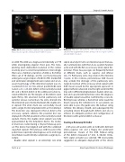
Video 22. Double interatrial septum. View supplemental video at http://dx.doi.org/10.12945/j.jshd.2016.005.16.vid.22.
an ASD. The ASD was diagnosed incidentally on TTE while investigating atypical chest pain. The trans- planting team demanded resolution of the cardiac anomaly prior to renal transplantation. Interestingly, there was a familial occurrence of ASD, as his father, three out of 10 siblings, and his son had previously undergone surgical repair of ASD. On TEE, marked RA and ventricular enlargement were noted, and an un- usual morphology of the interatrial septum was en- countered (Video 22). Two almost parallel ASDs were noted: a 23- × 28-mm defect in the normally located IAS and a 30-mm defect in the additional curtain lo- cated within the LA. The margins of the defects were quite imsy. We observed normal pulmonary and systemic venous connections. No veins drained into the interatrial space formed between the double atri- al septum. The atrial shunt was successfully closed with a single 38-mm Amplatzer ASO on rst attempt. The distal disc was deployed in the LA distal to the accessory septum, whereas the proximal disc was deployed in the RA proximal to the normally located septum. Hence, the double atrial septum was fully approximated by the Amplatzer device. No residu- al shunt was noted during a 10-year follow-up peri- od. The device had aligned well with the combined squashed septum. Percutaneous ASD closure in this patient was especially advantageous, as his end stage renal failure could critically complicate a surgical pro- cedure.
Double atrial septum is an extremely rare atrial
Video 23. Improper deployment of the entire device in the left atrium. View supplemental video at http://dx.doi.org/10.12945/j. jshd.2016.005.16.vid.23.
septal anomaly. It forms an interatrial space that usu- ally communicates with the LA via a patent foramen ovale and with the RA via accessory atrial septal fen- estration. These two passages are frequently formed at di erent levels, such as superior and inferior [26, 27]. Pulmonary veins may drain in the interatria l space; in this scenario, percutaneous ASD closure may occlude the drainage of this pulmonary vein. Surgical resection of the accessory atrial septum with ASD closure would be the appropriate approach. A pigtail catheter advanced into the right ventricle (RV) may aid in di erentiating between double atrial sep- tum and a prominent Eustachian valve; the diagnosis of double septum would be con rmed by a non-de- ecting tissue, whereas a Eustachian valve would be drawn away by the catheter [28]. In our patient, we were able to pass the guide wire, the balloon sizing catheter, the delivery sheath, and subsequently the occluding device through both defects and also to achieve an adequate position and con guration of the device with optimal defect occlusion.
Snaring a Runaway Occluder
A 65-year-old woman with an ASD with de cient antero-superior rim and a oppy IAS underwent percutaneous closure of the ASD. Balloon-sizing of the defect measured 27 mm. A 30-mm Occlu- Tech septal occluder was selected. TEE inaccurately suggested an adequate deployment of the device
Tal, R. et al.
Atrial Septal Defect Occlusion Challenges
