
Page 26 - Journal of Structural Heart Disease Volume 3, Issue 4
P. 26
113
Case Report
Figure 3. Transesophageal echocardiography in four-chamber view showing the perimemberanous ventricular septal defect partially covered by an aneurysm.
Figure 4. Transesophageal echocardiography in short axis show- ing good positioning of the ADO II.
With the introduction of Amplatzer devices, tran- scatheter closure of PM VSDs has become a well-es- tablished procedure but is associated with an unac- ceptable incidence of complete heart block [5]. The routine technique for percutaneous VSD closure is an antegrade approach accomplished by creating an ar- teriovenous loop. Here, we describe the case of a child who underwent percutaneous closure of a PM VSD using a retograde approach with an o -label ADO II in the presence of interrupted IVC and suitable anat-
El-Sisi, A. et al.
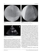
Figure 5. Cineangiography in LAO/60° cranial view showing the device completely deployed and released.
omy and diameter of PM VSD for device selection. In this situation, we believed that the transcatheter clo- sure of the PM VSD through internal jugular access would have been imprecise, increased the duration of the procedure, and ultimately been unsuccessful. However, the transcatheter closure of ASD and pat- ent ductus arteriosus has been established using an internal jugular approach in many previously report- ed cases of interrupted IVC [7, 8]. If the size of the PM VSD had been > 6 mm, we would not have been able to use the ADO II, as the maximum available waist diameter of the device is 6 mm. Therefore, we would have had two options: (1) referring the patient or (2) trialing the use of the azygous vein as alternative route for an antegrade approach. Using an antegrade approach to the percutaneous closure of a PM VSD in a patient with interrupted IVC was previously de- scribed by Kawar et al. [6]. Furthermore, our experi- ence with the percutaneous retrograde closure of a MP VSD with interventricular septum aneurysm using the ADO II is consistent with a report by Koneti et al. [2], who described successful retrograde approaches to PM VSD closure in a large group of 57 children with favorable anatomy.
In conclusion, interrupted IVC should be diag- Closure of VSD in Child with Interrupted IVC
